A guide to making lesion observations Disease outbreaks and mass mortalities of fish generate notable public concern and often require investigation on the part of fish heath diagnosticians and fishery managers. External lesions on fish are commonly observed in association with field morbidity or mortality events, and descriptions of these lesions can be helpful in diagnosing the cause of the etiologic agent(s), i.e., the cause of the event. However, the terminology used to describe external lesions is variable and may differ between agencies and individuals. Further, and most importantly, lesion observations are often non-specific with regard to the etiologic agent(s). This means that certain types of lesions can be caused by a variety of different bacteria, viruses, fungi or parasites, and you cannot tell by simply looking what the cause of a lesion is. For example ulcerative lesions can be associated with all of the aforementioned examples. Link here to see pictures of ulcerative lesions associated with different etiologies. With the support of the US Environmental Protection Agency, Office of Wetlands, Oceans and Watersheds, this guide to fish lesion observations was developed. A pdf version of this guide can be dowloaded by clicking here. Purpose of this lesion guide This guide was developed to present an overview of observation techniques and provide terminology that can accurately describe external fish lesions. You do not need to be a fish pathologist to make observations that can be used to make a diagnosis. Using a consistent approach, and with a little practice, field technicians, fisheries biologists and environmental managers can make relevant observations, take high quality tissue samples, and make substantial contributions to field fish health studies. To make good observations they should not only make sense to you, but the descriptions should not be misunderstood by others reviewing your data. Further, it is helpful to be systematic (i.e., follow a prepared, itemized "game plan" for making observations) and consistent (treat all observations and samples in the study the same way) in your sampling strategy. This guide was intended to facilitate observations both in the field as well as in the laboratory. This guide contains common terminology, sample descriptions and data sheets to assist with lesion observations. The data sheets have been provided for you to use "as is" or as a template to utilize data entry items or formats on your own data sheets. Good planning and use of appropriate data sheets plays an important role in determining the final outcome of your observations. Remember, you can never go back and get data that was not collected!!! Sections to this guide:
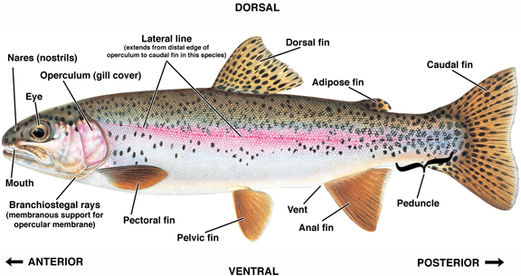
Although making visual observations of lesions does not necessarily involve direct, physical contact with animals, careful inspection or disposal of individual fish carcasses often does involve some form of contact. Therefore, it is prudent to use protective clothing to avoid contact with potential disease agents. Gloves, lab coat, apron, goggles, and other protection should be worn as needed. A list of vendors that supply these items is included at the end of this guide. When making collections or lesion observations each animal should have a unique ID number using a defined accession system. Useful information to include with your gross pathology observations includes: source of fish, water quality from source including temperature, source location, time and weather conditions during collection, collection method, general environmental observations, anomalous behavior (i.e., gulping at surface, flashing, morbidity, etc.), an estimate of the number of affected fish, relationship to previous reports, and nearby landmarks (i.e., marinas, sources of potential contamination, etc.). This section will allow you to clearly specify the anatomical location of any gross pathology observed. Examples of four fishes with varying anatomical features are annotated below for reference. These illustrations were contributed, with permission, by Joseph Tomelleri (Cimmeron Trading Co. http://www.americanfishes.com). These images may not be reproduced, used or distributed except as part of this intact outreach guide to fish lesions. The first fish image shown, rainbow trout, is annotated with dorsal-ventral and anterior-posterior orientation:
External anatomy of the rainbow trout (Oncorhynchus mykiss). Fish illustration by Joseph Tomelleri (with permission), general annotation by A. Kane.
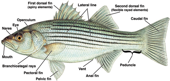
External anatomy of the striped bass (Morone saxatilis). Fish illustration by Joseph Tomelleri (with permission), general annotation by A. Kane.
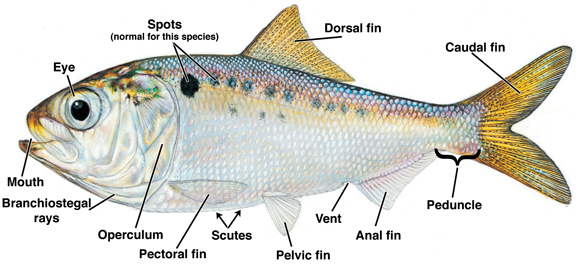
External anatomy of the gulf menhaden (Brevoortia patronus). Fish illustration by Joseph Tomelleri (with permission), general annotation by A. Kane.
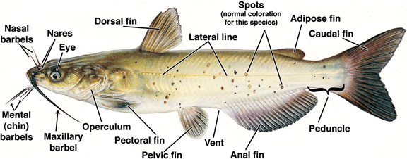
External anatomy of the channel catfish (Ictalurus puncatus). Fish illustration by Joseph Tomelleri (with permission), general annotation by A. Kane.
External Examination (checklist of parameters that may be relevant for comment):
Good lesion observations are descriptive and systematic. Record detailed observations of any lesion or area that may be abnormal. In any samples taken for histopathology, include pieces of the abnormal tissue with a bit of surrounding, apparently normal tissue. To provide maximal imagery of your observations, include comments on:
Use the above comment categories and "food descriptions" to convey the lesion imagery, particularly when the anomaly which you are describing is unknown to you. Examples of material or food imagery which can be used to describe lesions include: cauliflower, cottage cheese, cream cheese, pea soup, cream and coffee, marbles, sandpaper, etc. Imagery can also be used for size comparisons if no ruler is available, e.g., size of a pin head, pencil eraser, nickel, dime, quarter, orange, basketball, etc. Please refer to the simple lesion classification system below for additional information. Further, we suggest that you draw a picture or use data sheets that contain a fish line drawing and draw in the abnormality indicating location. Complement the drawing with descriptive words. Sample data sheets are provided so that you can record lesion observation data:
A Simple lesion classification system: 1. LESION OBSERVATION (describe primary lesion first; make additional observations for other lesions):
2. LESION LOCATION (e.g., head, vent, left pectoral fin, lateral line, etc.) 3. LESION PRESENTATION
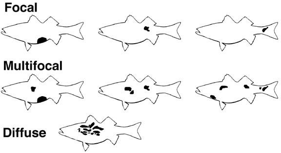
4. LESION SEVERITY : May be ranked on a scale of 1-3 or 1-5; 0=no lesion. If using the 0-3 scale, the mid-range severity ("moderate," with a rank of 2) covers a broader range of severity than mild or severe in the 0-5 scale. See diagram below. The 0-5 scale affords greater range of observation. Cartoon illustrating different lesion presentations:
Additional Examination Techniques: Systematic necropsy : Although necropsy techniques are outside the scope of this guide, we can mention that non-professionals can learn to sample all organ systems for preservation and subsequent histological examination. Several references are provided at the end of this guide that include necropsy techniques. All organs should be sampled for necropsy, regardless of the presence of grossly observable lesions. Organs should include gills; heart with ventricle, atrium and bulbus arteriosis; skin; muscle; bone/cartiledge; brain; eye; lateral line; esophagus; stomach; intestine; anterior kidney; posterior kidney; swim bladder; liver; spleen; mesentery and gonad.
Tissue sampling and preservation techniques for histopathology:
Recipe for 10% Neutral Buffered formalin (to make 1 liter):
Safe and proper sample shipment and cleanup:
Photographic documentation of lesion observations If possible take a photograph (note photograph number of the lesion/specimen). Include a ruler (or common object, e.g. a coin) in the picture to indicate relative size. Photographs provide the pathologist with accuracy and the greatest possible information). a) practice in advance - take test pictures with exposure bracketing; Thanks to "modern times," digital photography has become user-friendly and affordable. Use of digital photography and a computer with internet accessibility, allows fish culturists and other persons to take digital pictures of whole fish, lesions and microscopic images, and forward them to colleagues or fish health experts for evaluation. Low-end, but highly utilitarian digital cameras cost between $150 and $350. If you go this way, be sure to have "macro ability," i.e., the ability to photograph small objects like lesions at 1:1 (where a dime could fill the image frame). If you are already in this "digital age," you can share your observations with others easily over the internet! This concept can be a valuable tool to send observation data to a fish heath manager or pathologist.
Sample lesion observations:
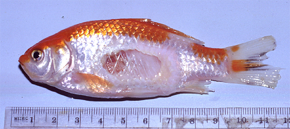
This goldfish (Carassius auratus) has a focal, well-circumscribed, deep oval ulcer (severity ranking of 5, "severe"), 25 x 18 mm, located on the left mid-flank, above the pelvic fin. The ulcer penetrates through the muscle and reveals rib bones and some of the viscera. The margins of this ulcer do not appear reddened, but the posterior margin has a cream-colored section extending approximately 3-4 mm. The distal edges of the anal and caudal fins are frayed (serverity ranking of 3, "moderate").
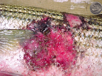
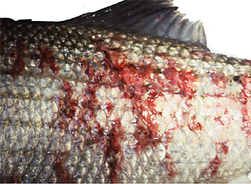
The image of the striped bass (Morone saxatilis) on the left has a large (7 x 6 cm), focal, diffuse irregularly-shaped, red lesion (ulcer) that penetrates the skin and scales and reveals underlying dermis (severity ranking 4, "marked"). The lesion is located on the left side of the specimen, extending posteriorly from the distal edge of the pectoral fin; a portion of the lesion extend to the dorsal surface of the fish, anterior to the first dorsal fin. Portions of the lesion, as well as the lesion margins, have superficial black coloration (possibly pigmentation). The lesion margins are irregular. The surface of the lesion has a sandpaper-like appearance. Ventral to the lesion, there is a large, diffuse area of reddening, with no loss of scales, that extends from the branchiostegal membrane to the vent. The fish was in otherwise good body condition. In contrast, the image of the striped bass on the right shows a multifocal, extensively distributed lesion.
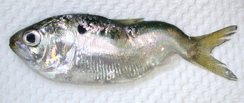
This young-of-the-year Atlantic menhaden (Brevoortia tyrannus) measured 58 mm total length and presented with spinal deformity. The deformity consisted of curvature in the dorsal-ventral plane, and extended from the middle of the dorsal fin to the caudal fin. Severity ranking 3, "moderate." The animal was alive and otherwise non-remarkable when observed.
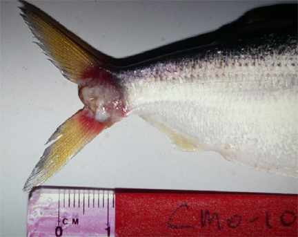
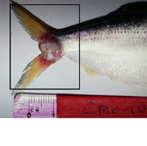
This specimen is an Atlantic menhaden (Brevoortia tyrannus) that presented with a raised, irregularly-shaped, off-white, cauliflower-like growth (5 x 8 mm) on the right side, extending from the peduncle into the fork of the caudal fin. The mass has well-defined edges and had reddened margins. The smaller photograph on the right shows the same image and indicates the appropriate size tissue sample that would be taken for preservation and subsequent histologic analysis. This sample contains both the abnormal tissue as well as adjacent normal tissue.
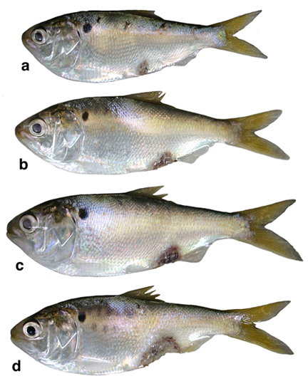
This composite image of four young-of-the-year Atlantic menhaden ( Brevoortia tyrannus ) depicts ulcerative lesions with varying ranks of severity. The ulcer seen on fish "a" has a severity ranking of 2, "mild." Fish "b," "c" and "d" have severity rankings of 3 (moderate), 4 (marked) and 5 (severe), respectively. Of course these rankings are relative and subject to the experience of the observer. Nevertheless, if personnel follow the basic tenets of severity rankings as illustrated in this guide, there should be a fair degree of consistency between observers, plus or minus one severity ranking. Note that the ulcer on fish "a" is located halfway between the pectoral fin and the anal fin, whereas ulcers on the other three specimens are perianal. These perianal lesions have burgundy margins with central red to grayish areas. Also, careful observation of the image of fish "a" shows a small (3 x 3 mm), diffuse area of reddening on the left side, just below the distal insertion of the dorsal fin. AFS (American Fisheries Society) 1992. Investigation and Valuation of Fish Kills. American Fisheries Society Special Publication 24, Bethesda, Maryland. Austin, B. and D.A. Austin. Bacterial Fish Pathogens. John Wiley and Sons, NY 1987. ASTM (American Society for Testing and Materials ) 1996. Standard Guide for Fish and Wildlife Incident Monitoring and Reporting. ASTM E1849-96. Ferguson, H.W. Systemic pathology of fish. Iowa State University Press. Ames, Iowa, 1989. Gratzek, J.B., Matthews, J.R. Aquariology. The science of fish health management. Tetra Press. Morris Plains, NJ, 1992. Kane, A.S., ed., FishGuts, A Multimedia Guide to the Art and Science of Fish Anatomy, Health and Necropsy on CD-ROM for Mac/Win. APC Press, 1996. Noga, E. Fish Disease Diagnosis and Treatment. Iowa State University Press. Ames, Iowa, 2000. Reimschuessel, R., Bennett, R.O., Lipsky, M.M. A Classification system for histological lesions. J. Aquat. Anim. Health; 4: 135-143, 1992. Reimschuessel, R., May, E.B., Bennett, R.O., Lipsky M.M. Necropsy examination of fish. Veterinary Clinics of North America: Small Animal Practice; 18:427-433, 1988. Ribelin, W.E., Migaki, G. eds., The pathology of fishes. The University of Wisconsin Press. 1975. Roberts, R.J. ed., Fish Pathology. 2nd. Ed. Bailliere Tindall. London, England, 1989. Stoskopf, M. Fish Medicine. W.B. Saunders Company, Philadelphia, P.A., 1993. Ordering Information for Fish Necropsy Supplies You can request a catalog from these and other companies so that when you need to make budgeting decisions and/or order supplies, the information is at your fingertips. Mention of suppliers in this guide does not constitute endorsement. Argent Chemical Laboratories - MS222 and other chemicals/drugs
Aquatic Eco-Systems - Aquaculture hardware. Great catalog
Biomedical Research Instruments, Inc. - medical and dissection instruments
Fisher Scientific - General scientific supplies
IDE Interstate Inc. - medical equipment and instruments, pharmaceuticals and proprietaries
Thomas Scientific - General scientific supplies
VWR Scientific Products - General scientific supplies
Website and content maintained by UF Aquatic Pathobiology Laboratory
|
|||||
|