INTEGUMENTARY SYSTEM Description
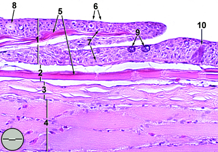
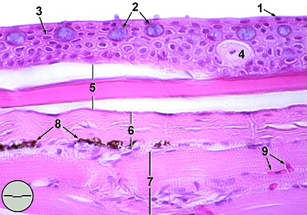
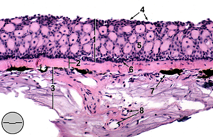
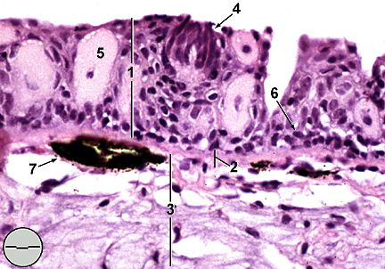
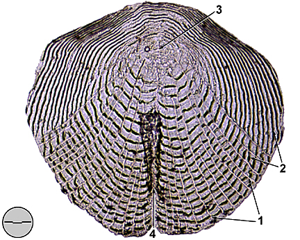
The integumentary system or skin of the fathead minnow is composed of two layers, an outer epidermis and an underlying dermis (Fig. 1). The epidermis is additionally divided into an outer fusiform layer composed of stratified squamous epithelial cells and a basal layer (stratum basale) of undifferentiated germinal cells. In general, thickness of the epidermis is greatest on the dorsal surface and toward the head (10-15 cells) and thins caudoventrally (2-3 cells). The dermis is composed primarily of collagenous connective tissue distinguished as stratum compactum, a dense collagenous matrix providing structural strength and stratum spongiosum, a loose network of collagen and reticulin fibers. Dermis also contains pigment cells called chromatophores. Other cells found within the epidermis include; mucous cells, alarm cells , and taste buds . Mucous cells derive from undifferentiated basal cells as they increase in size and migrate to the surface(Fig. 2). Mucus secretions, composed primarily of glycoproteins, form a slimy protective coat. Functions attributed to this coat include drag reduction (Hoyt, 1975), predator evasion, and isolation of surficial epithelial cells from bacteria. Immunoglobulins, also present in mucus, provide additional protection against infection. Alarm cells, found in greatest densities on the head, release fright substances (Schreckstoff) when ruptured (Fig. 3). Upon detection these substances incite predator avoidance responses in other fatheads and may serve to reduce cannibalism (Moyle and Cech, 1996). Cutaneous taste buds, also found predominantly on the head and ventral surface, are chemoreceptive and assist in locationg food (Fig. 4). Typically taste buds are bulb shaped structures comprised of sensory, basal, sustentacular, and marginal cells. They emanate from the basement membrane often protruding above the epithelial surface. Cutaneous taste buds are innervated by the branches of the facial nerve (cranial nerve VII). Prominence of the facial lobe of the medulla oblongata indicates the reliance fathead minnows place on gustatory sensation (Kapoor, et al., 1975). Fathead minnows are covered with an armor of cycloid scales (except on their heads which, like all Cyprinidae, are scaleless). Cycloid scales are translucent, acellular plates of collagenous tissue which anchor within dermal scale pockets in the stratum spongiosum but project through the basement membrane into the epidermis (Fig. 5). They are covered by a thin squamous epithelium which remains distinct from epidermal tissue. Scales overlap one-another in an imbricated manner with free ends directed caudad. They tend to be of moderate size but are much reduced and irregularly arranged in the predorsal region (between head and dorsal fin)(Jenkins & Burkhead, 1994). Breeding male fathead minnows develop horny nuptial tubercles
and a spongy nape pad. Tubercles are hard (karatenized), conical
structures which derive from epidermal cells. As many as 24 large
tubercles may form in several rows on the anterior and internasal
regions of the snout. Tubercles function in aggression between
males, sexual stimulation of females, and maintenance of spawning
sites (Wiley and Collette, 1970). The nape pad develops on and
covers the dorsal surface from the margin of the head to the dorsal
fin. This specialized pad contains mucous-secreting cells and
taste buds. Suggested function of the nape pad include marking
of spawning sites, chemosensory assessment of egg condition, and
fungicidal protection of egg masses (Jenkins & Burkhead, 1994).
INTEGUMENTARY
SYSTEM: Caudal peduncle, longitudinal section (a)
INTEGUMENTARY
SYSTEM: Caudal peduncle, longitudinal section (b)
INTEGUMENTARY
SYSTEM: Integument of the head, transverse section (a)
INTEGUMENTARY
SYSTEM: Integument of the head, transverse section (b)
INTEGUMENTARY
SYSTEM: Scale
[Back to Table of Contents]
|