REPRODUCTIVE SYSTEM: Male Description
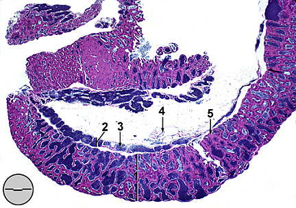
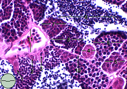
DESCRIPTION The fathead minnow testis, like the ovary, has bilateral elongate lobes located within the abdominal cavity and suspended from the gas bladder by mesenteries. These mesenteries, termed mesorchia, encase a thin tunica albuginea which, in turn, encases the testicular lobes. Each lobe is comprised of a longitudinally oriented collecting duct (ductus deferens) along the length of which radiate seminiferous tubules (Fig. 1). The thin capsular walls of these tubules are formed by invagination of the tunica albuginea. Spermatogenesis, the maturation of germ cells to spermatozoa, takes place within the seminiferous tubules. Following maturation sperm are stored in tubule lumena and within the ductus deferens. Caudally the ductus deferens from each lobe merge before exiting at the genital orifice located between anus and urinary pore. Spermatogenesis is said to be unrestricted in fathead minnows meaning that maturation of sperm occurs along the entire length of the seminiferous tubules (Grier, 1981). Germ cells progress through six distinct cytological stage during spermatogenesis; primary spermatogonia, secondary spermatogonia, primary spermatocyte, secondary spermatocyte, spermatid, and spermatozoa (Fig. 2). Germ cells located within the stroma of the tubule wall give rise to primary spermatogonia. These are large cells with eosinophilic cytoplasm and a distinct nucleus containing dense chromatin. Each primary spermatogonia undergoes a series of mitotic divisions to produce a cluster of secondary spermatogonia encapsulated within a cyst. The cyst arises from a Sertoli cell associated with the original germ cell. Clusters of cells resulting from divisions of the original germ cell maintain a consistent stage of development within the cyst Secondary spermatogonia are smaller than primary spermatogonia with large lightly basophilic nuclei and little cytoplasm. Primary spermatocytes, the result of another round of mitotic divisions, are smaller still with increasingly basophilic nuclei. Primary spermatocytes undergo the first meiotic division to produce secondary spermatocytes. Still contained within the cyst, these cells are again smaller and have increasingly dense basophilic nuclei. Secondary spermatocytes undergo a second meiotic division. The resulting spermatids have condensed intensely basophilic nuclei and very little cytoplasm. At this stage the cyst ruptures releasing the spermatids into the lumen where final maturation takes place. Spermatids Each spermatid develops into a spermatozoan.
REPRODUCTIVE
SYSTEM: Mature testis, sagittal section
REPRODUCTIVE
SYSTEM: Seminiferous tubule, transverse section
|