REPRODUCTIVE SYSTEM: Female Description
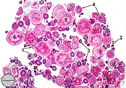
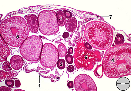
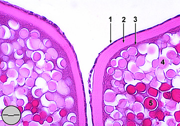
Fathead minnow ovaries are bilateral elongate lobes oriented longitudinally within the abdominal cavity and containing ova at varying stages of maturity (Fig. 1). They suspend from the ventral surface of the gas bladder by mesenteries termed mesovaria (Harder, 1975). Arteries, veins, lymph vessels, and nerves that supply the ovaries enter via the mesovaria. The ovaries themselves are enclosed in a fibrous connective tissue tunica albuginea which is contiguous with the mesovaria (Fig. 2). The lumenal surface of the tunica albuginea folds into ovigerous lamellae oriented perpendicular to the long axes of each ovarian lobe. Lamellar walls are composed of germinal and follicular epithelia supported by a connective tissue stroma. Development of ova from germinal cell to mature egg is divided
into six stages defined according to morphological characteristics
of nucleus, oviplasm, and follicular wall. Oocytes increase in
size as they proceed through the developmental stages. Mitotic
proliferation of germinal cells produces clustered of Stage I
oocytes (oogonia) nested within the germinal epithelium. These
cells have large nuclei and lightly eosinophilic cytoplasm. Stage
II oocytes have basophilic cytoplasm and a conspicuous central
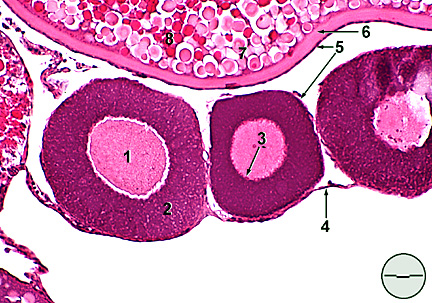
nucleus with diffuse chromatin. Stage III oocytes break from the
germinal epithelium continuing maturation within the folds of
the ovigerous lamellae. At this stage a simple squamous follicular
epithelium envelopes the ova and provitelline nucleoli become
evident in the karyoplasm (Fig. 3).
Stage IV is defined by the appearance of yolk granules and fat
vacuoles in the oviplasm. Euvitelline nucleoli are evident along
the nuclear membrane and a distinct vitelline envelope (chorion)
appears beneath the follicular epithelium. Yolk vesicles increase
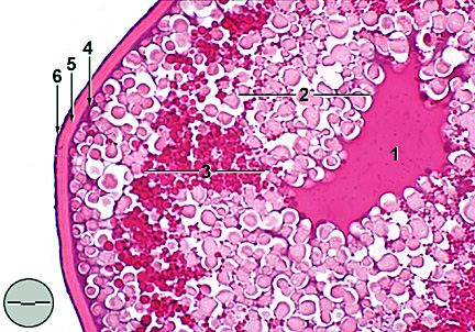
significantly during stage V filling the entire oviplasm except
narrow basophilic bands around the nucleus and beneath the vitelline
envelope (Figs. 4 & 5).
The nucleus contains fewer uvitelline nucleoli and the nuclear
membrane begins to degenerate. Prior to final maturation the nucleus
migrates peripherally
REPRODUCTIVE
SYSTEM: Ovary (a)
REPRODUCTIVE
SYSTEM: Ovary (b)
REPRODUCTIVE
SYSTEM: Stage III oocytes
REPRODUCTIVE
SYSTEM: Stage V vitellogenic oocyte
REPRODUCTIVE SYSTEM: Stage V oocytes,
detail of the follicular wall
|