|
Case History
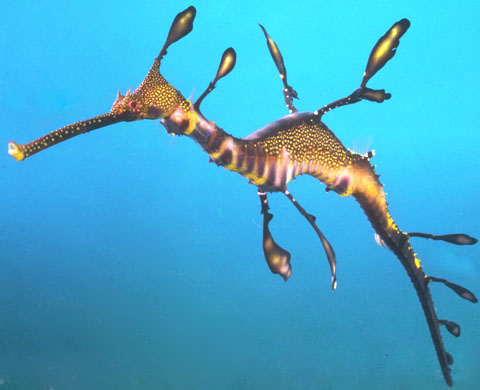
Four weedy Sea Dragons in tank; within 2 day period, two animals were found in lateral recumbency at the bottom of the tank. They had high respiratory rates and skin appeared to be sloughing off the body. Treatment with Cicrofloxicin and liquid B vitamins was initiated. The animals became apparently stable with decreased respiratory rates, but did not improve further. Two days after treatment, a third animal became moribund and was submitted for diagnostic evaluation. |
|
Physical Examination
The animal is seen here in lateral recumbancy. Prior to removing from transport bag, this animal was observed to have rapid restpiration rate.
|
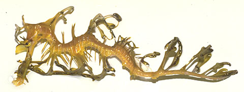
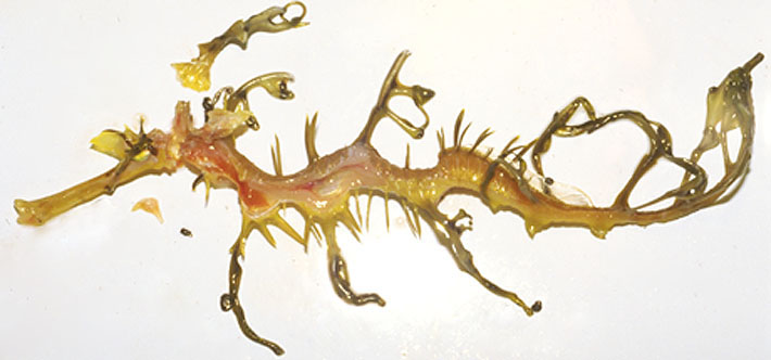
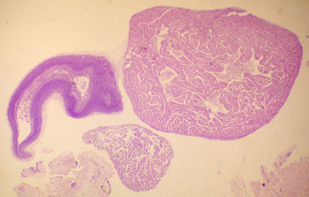
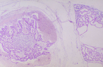
Gross Examination
Observable here are the organs in situ. The heart, not easily observed in this photograph, is just caudal to the gill region. There was no food evident in the intestinal tract; wet mount from a gut scraping was not remarkable.
Roll your mouse cursor over the image to identify the reddish-brown liver, the intestine, swim bladder and spleen.
|
Gills
|
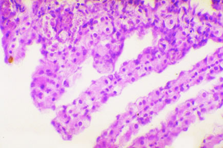
Histopathology
Brain and eyes- (not shown) were not- remarkable.
Gills: note the epithelial cell hypertrophy and necrosis
Roll your mouse cursor over the image to see these areas.
Red arrows indicate hypertrophy, blue arrows indicate necrosis. |
|
|
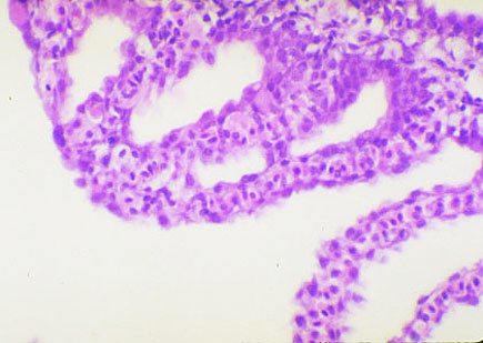
Histopathology
More Gills
In some foci there is fusion of the secondary lamellae tips. Note the frayed appearance of the respiratory epithelium undergoing degenerative changes.
|
Cardiovascular system
|
Histopathology
Cardiovascular system
The phagocytic endothelium in all of these structures is hypertrophic and hyperplastic .
This is even evident at low magnification in the lumen of the bulbus arteriosis. Two higher magnifications of the bulbus arteriosis are available below.
Roll your mouse cursor over the picture to identify these structures. |
| |
Histopathology
Bulbous Anteriosis
Medium Magnification (20x)
The endothelium should cnsist of a single layer of flat cells with linear basophilic nuclei and little to no cytoplasm. As you can see here, the endothelial cells contain a large amount of eosinophilic cytoplasm. There are multiple layers of endotheial cells extending into the lumen, almost obstructing the luman in some areas. Note the basophilic elastic fibers in the media of the bulbus arteriosis.
|
|
|
Histopathology
Bulbous Anteriosis
Medium Magnification (40x)
Hypertrophy and hyperplasia of the phagocytic endothelium , which is a part of the teleost reticuloendothelial system, is a response frequently found in fish with systemic infections. |
|
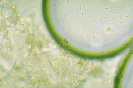
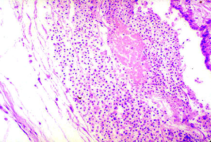
Histolopathology
Scrape contained numerous tear- shaped ciliates that were between 30-40 uM in length with a prominent. more pointy anterior end. One of these organisms is seen in this wet pount trapped on the periphery of an air bubble. Wet mount of gill biopsy revealed numerous motile, rod- shaped bacteria and occasional flagellates (8- 10uM); photo not available.
|
|
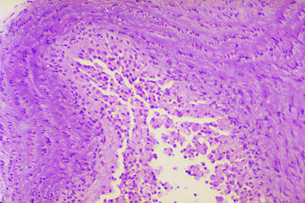
Histolopathology
Skin, Low Magnification (4x)
Most of the overlying epithelium has undergone necrosis. Note the pigment containing cells in the outer layer the dermis, and the dark stained parasites below. |
|
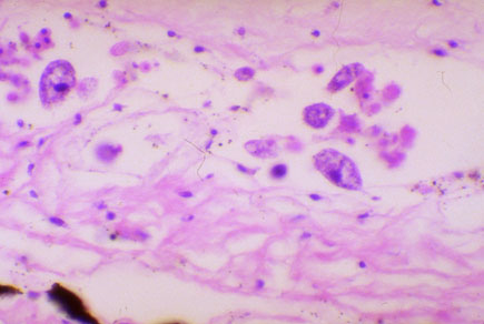
Histopathology
Skin
High Magnification (40x)
In this high magnification of the dermis, there are multiple protozoan parasites in the connective tissue and within dermal blood vessels.
|
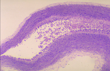
Swim Bladder Rete and
Posterior Kidney
|
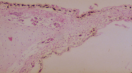
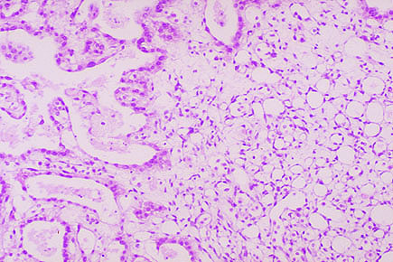
Histopathology
Swim Bladder Rete and
Posterior Kidney
Low Magnification (4x)
Here you can see the extensive dilation of the renal tubules in the posterior kidney . Ventral to the kidney is an area of loose connective tissue and the swim bladder. Note the prominent rete capillary network and the large area of hemorrhage.
Roll your mouse cursor over the micrograph to identify these stuctures. For a higher magnification of the structures, click on them with the mouse.
|
|
Capillary Network
(rete)Swim Bladder
The rete provides an extensive network of capillaries for gas exchange between the blood and swim bladder.
|
|
Area of Hemorrhage
A higher magnification of another focus of hemorrhage shows a large eosinophilic area of necrosis surrounded by degenerating red blood cells. Present also is edema surrounding the loose connective tissue adjacent to the swim bladder
|
|
Posterior Kidney
The renal tubules are extremely dilated and there is moderate edema of the interstitium. These findings are compatible with acute tubular necrosis and renal failure. Fish in renal failure have difficulty maintaining normal osmoregulation. Notably absent from this kidney are glomeruli. These fish are similar to the seahorses, which also are aglomerular.
|
|
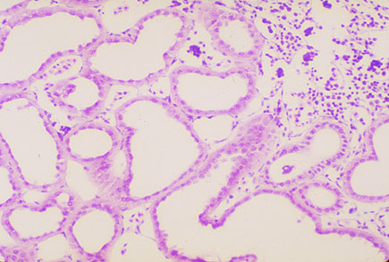
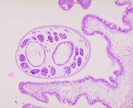
Histopathology
Intestine
High Magnification (40x)
This section of intestine shows a cross section of a digenetic trematode in the lumen. This parasite has a bifurcated gut (the two large vacuolar structures,) and a large amount of gonadal tissue (the basophilic tissue in the periphery of the parasite).
The adjacent intestinal mucosa is intact in this section. Note the clear mucous cells present in the intestinal epithelium. No evidence of inflammation is present in the mucosa or submucosa. This parasite is an incidental finding, and was probably not a factor in this animal's demise. Parasitism is frequently found in wild caught specimens, and is often the norm rather than the exception.
|
Diagnosis:
Cause of death: euthanasia
Heart: atrium, phagocytic endothelial cell hypertrophy, moderate, diffuse.
Bulbus arteriosis: endothelium, hypertrophy, hyperplasia, severe, diffuse.
Gills: epithelial cell necrosis, marked, diffuse.
Gills: epithelial cell hypertrophy, moderate, diffuse.
Intestines: parasitism, chronic, moderate, multifocal.
Peritoneum: peritonitis, chronic, moderate, multifocal.
Spleen: splenitis, chronic, moderate, multifocal.
Kidney: tubular dilation, marked, diffuse.
Kidney: interstitial edema, marked, diffuse.
Kidney: acute tubular necrosis.
Skin: dermatitis, ulcerative, parasitic, protozoan, supporative (definable button: "associated with the generation of pus; inflammation caused by the presence of pyogenic (pus-forming) bacteria), severe, diffuse.
Comments: The protozoan parasites noted on gross postmortem caused extensive epidermal ulceration, which in turn caused osmotic failure and acute tubular necrosis, with possible migration through other host tissue. Parasitological evaluation of the parasites using special stains reveals these ciliates to be Uronema. Had this animal not been euthanized, cause of death would most likely have been failure of osmotic regulation. For additional information, use reference texts to learn more about effects of protozoans on vasculature and release of cytokines.
|
|
|