| |
Case History
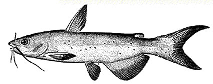
Mr. Joe Morgan, owner of Joe's Pond Fishin', complained of chronic mortality of channle catfish in his small rearing ponds in early spring. Several animals were evaluated on-site by external examination. They appeared emaciated. Mr. Morgan suggested treating his ponds with formaldehyde to control parasites.
|
| |
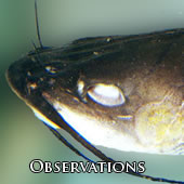
Initial Observations:
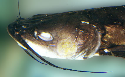
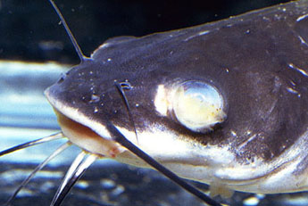
Animals exhibited "big head syndrome," i.e., they were emaciated. Several of these animals were so thin that they had sunken eyes. The catfish in this picture also shows a circular, yellowish 1 cm lesion on the right preoperculum. This is probably a bacterial lesion and should be cultured (along with the liver or posterior kidney). |
More Observations:
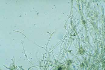
Below, a fish from the same pond exhibited a whitish yellow fuzzy coating over the left eye, and some reddening around the mouth and chin. The image on the left is a 10x magnification of this wet mount. It shows numerous branching, non-septate fungal hyphae. |
| |
Further Observations
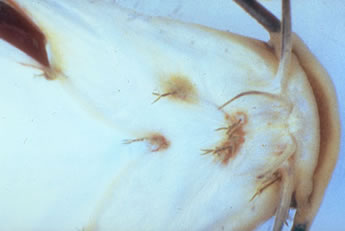
Observable on the chin, isthmus, and branchiostegals, are numerous copepods embedded into the skin. There is reddening (inflammation) around the site of attachment to the host.
|
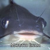
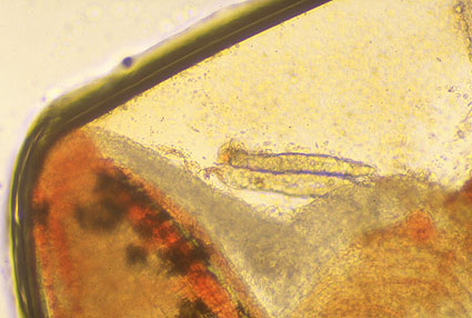
Mouth Exam
Upon examining the oral cavity of this animal, we observe a copepod attached to the tongue. It is put into some pond water until we can get it to a dissecting microscope.
|
|
Copepod Taxonomy
Kingdom:
Phylum:
Subphylum:
Class:
Order:
Family:
Genus: |
Animalia
Arthropoda
Crustacea
Copepoda
Lerneopodidea
Lerneopodidae
Achtheres
|
|
Parasitic Copepo
You are looking at the anterior end of this copepod. On the left is the small, mushroom-shaped organ of attachment, the bulla. Two arm-like structures connect the bulla with the head (or cephalothorax). These arms are actually modified second maxillae. The cephalothorax is connected by a narrow neck region to the animal's trunk. Extending from the trunk are the conspicuous elongate, cylindrical egg sacs.
It is interesting to note that this parasite is permanently secured within the tissue of the fish by insertion of the bulla. Ingestion, however, is accomplished through a small mouth seen at the top of the head (underneath the arms).
|
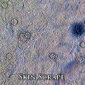
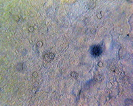
A skin scrape and gill biopsy were preformed on several fish from the affected pond. A microscopic view of the skin scrape may be seen below
To the right, the image at 10x magnification, numerous (approximately 14) found parasites can be observed in a single field. |
Skin Scrape
Low Magnification
|
The video below of a Higher magnification (40x) reveals these ciliated parasites to be Trichodina sp. Nothe the wreathes of beating, locomotory cilia around the periphery of the parasite, and the more central ring of interlocking denticles.
|
Skin Scrape
High Magnification
Kingdom:
Phylum:
Class:
Order:
Family:
Genus: |
Protista
Ciliophora
Ologohymenophorea
Mobilina
Trichodinidae
Trichodonia
|
|
|
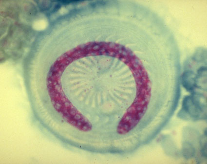
| Use of Feulgen stain emphasizes both the large horseshoe-shaped macronucleus, and the smaller micronucleus. The presence of cilia, and two different kinds of nuclei, are key features of ciliate protozoans. |
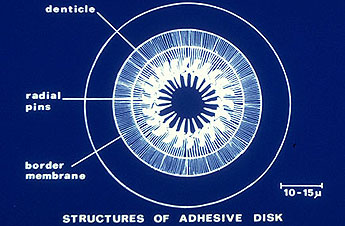
View of aboral (ventral) surface showing interlocking dentices, radial pins and border mambrane. These structures on the parasite work in unison to contract and cause suction to attach to the fish host. |
|
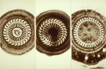
Silver Nitrate staining of the parasite allows us to observe some of the beautiful variation of denticle patterns within the family of Trichodinides. |
Another Skin Scrape
The video to the left is a different skin scrape from another channel catfish. Although difficult to see at this low magnification, there is a small protozoan in the center of the screen. By its movement, it is probable a ciliate.
The video on the right shows a higher maginification(1000x), this single-celled parasite is definitely 'Ich.' What gives it away is the spherical shape, horseshoe shaped macronucleus, and the slow, rotational movement. This movement is facilitated by holotrichous [definalbe button] cilia, not easily seen under lighting conditions in this movie.
|
|
Ichthophthirius Multifiliis
Kingdom:
Phylum:
Class:
Subclass:
Order:
Family:
Genus:
Species |
Protista
Ciliophora
Ologohymenophorea
Hymenostomatia
Hymenostomatida
Ichthyophthiriidae
Ichthyophthirius
Multifiliis
|
|
|
|
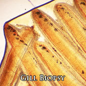
Gill Biopsy
Wet Mount
This is a wet mount from a gill biopsy. Note the long primary lamellae. There is one parasite observable in this picture. With only one parasite in the low magnification microscope field, the trematode infestation here would be considered mild. Compare this to the next mircograph.
|
Mild Infestation
With only one parasite in the low magnification microscope field with several lamellae, the trematode infestation would be considered mild. Compare this to the micograph at the bottom of the page.
|
Trematode
High Magnification
At higher magnification (40x), you can see a trematode attached to this primary gill lamellus.
|
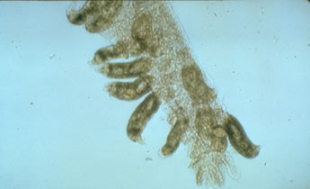
Trematode
This monogenetic trematode is very active, and is attached to the host gill tissue by its posterior end.
Kingdom:
Phylum:
Class:
Subclass:
Suborder:
|
Animalia
Platyhelmenthyes
Trematoda
Monogenea
Monopisthocotylea
|
|
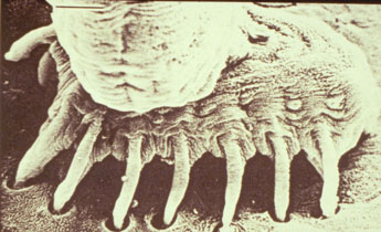
SEM Image
This scanning electon micrograph, magnified approximately 75,000x, shows the marginal hooklets of the haptor embedded into the
surface of fish host tissue. This type of damage
to host epithelium may result in secondary
bacterial infection.
|
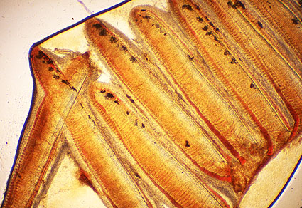
Severe Infestation
Over ten trematodes are evident on this one gill lamellus. If this gill filament were representative of others, the parasitic infestation would be considered severe.
Ranking system:
0- Non-remarkable
1- Minimal
2- Mild
3- Moderate
4- Marked
5- Severe |
Preliminary case report: #407-93
Client: Joe's Pond Fishin', Alabaster, New York; (607) 555-4567
Species: Channel catfish, Ictlurus punctatus
Date: 12 April 1993
Mr. Joe Morgan, propriator, requested on-site evaluation of chronic mortalities in 2 of his catfish ponds. Upon site visit, we noted that there was a large amount of organic debris in the ponds. Water quality parameters: pond temperature = 11 degrees C; air temperature = 16 degrees C; bright sunny day; dissolved oxygen (taken from row boat at center of pond; depth of 2 feet, 2:30pm) = 6.2 mg/L; mean depth reported to be 2.1 feet; pH 6.8.
Four animals were captured by hook and line, and brought back to the Fish Health Laboratory for external evaluation. Examination revealed these fish to be channel catfish, weighing 68-124 grams; 114-162 mm fork length.
skin, parasitism, marked, multifocal, Trichodina.
skin, parasitism, mild, multfocal, ichthyophthirius
skin, dermatitis, hemorrhagic, parasitism, marked, multifocal, copepods
skin, dermatitis, supperative, necrotizing, marked, focal
eye, cornea, keratitis, ulcerative, mycotic, marked, diffuse
gills, parasitism, mild, multifocal, monogenetic trematodes
tongue, parasitism, mild, focal, copepod
The general body condition of these fish indicated that they were emaciated. One animal was opened, and there was no abdominal fat evident. General pond observations and discussions with pond owner indicated that pond stocking densities are too high, and ponds have a significant amount of organic debris. This may cause a high BOD with oxygen deficits. The high stocking density and organic content of the ponds, along with the predisposition of poor condition, make the parasitism favorable. Pond treatment with formaldehyde will most likely result in significant mortalities. If feasible, suggest draining ponds and redigging to remove the years of organic debris; restock and monitor fish health, feeding and condition closely. |
|
|