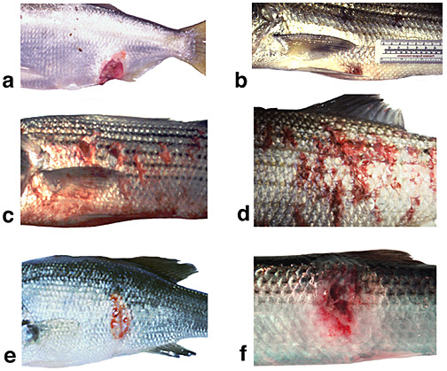
Ulcers can be present as small, discrete lesions or larger, more diffuse areas of reddening with loss of epithelium, dermis and underlying musculature. This panel of images shows examples of ulcerative lesions associated with different etiologies (causes). Panel a: ulcer on a menhaden associated with an oomycete fungus. Panel b: Small focal ulcer on a striped bass associated with Mycobacterium. Panel c: Diffuse areas of reddening and multifocal dermal ulcers on a striped bass infected with Mycobacterium. Panel d: Shallow, diffuse ulceration in a striped bass associated with the bacterium Edwardsella. Panel e: Focal ulcer on a largemouth bass associated with a microsporidian parasite infection. Panel f: Ulcer on a Pacific herring associated with viral septicemia virus and organic pollution. The take home message from these images is that ulcers are multifactorial in nature and non-specific with regard to the different environmental stressors that may cause them. Further, the presentation of an ulcer does not serve as an accurate telltale of the extent of internal symptoms or pathology. |
Website and content maintained by UF Aquatic Pathobiology Laboratory
and the Emerging Pathogens Insitute