DIGESTIVE SYSTEM: Liver
and Pancreas
Liver:
Description
Figure 1. Anterior abdominal cavity,
transverse section
Figure 2. Cords of hepatocytes and
central vein, transverse section
Figure 3. Sinusoids and bile ducts
Figure 4. Sinusoids and bile canuliculi
Pancreas:
Description
Figure 5. Exocrine and endocrine
tissue (a)
Figure 6. Exocrine and endocrine
tissue (b)
LIVER: Description
The liver is a largest of the extramural organs. It is roughly
U-shaped, situated ventral to the esophagus and conforming to
the peritoneal cavity and surrounding viscera. The color varies
from dark brown to cream or even yellow. Functions of the liver
include assimilation of nutrients, production of bile, detoxification,
hematopoiesis, and effete red cell destruction.
Parenchyma of the liver is contained within a thin capsule of
fibroconnective tissue. Often the capsule is not distinguishable
in light microscopic preparations. The parenchyma itself is primarily
composed of polyhedral hepatocytes typically with central nuclei.
Vacuolization of hepatocytes resulting from glycogen and/or fat
storage can produce considerable histological variability. Other
cell types typically found in liver parenchyma include hematopoietic
tissue and macrophage aggregates.
Venous blood enters the liver caudally from the intestine via
the hepatic portal veins and branches into capillaries known as
sinusoids. After passing through the sinusoids and collecting
in central veins the blood exits the liver via the hepatic veins
eventually returning to the heart via the sinus venosus. Sinusoids
are lined with reticuloendothelial cells which are in turn lined
with hepatocytes. Adjacent sinusoids are separated from one-another
by at least two hepatocytes. In the case of glycogen vacuolization,
the nuclei and cytoplasm of hepatocytes are compressed eccentrically
toward the sinusoidal spaces.
Bile ducts also occur within the parenchyma of the liver. Originating
between adjacent hepatocytes, bile canaliculi anastomose to produce
ducts of increasing diameter. Eventually the ducts merge to form
the common bile duct. Smaller ducts within the liver are lined
with a single layer of cuboidal epithelial cells. Larger ducts
may incorporate a layer of connective tissue and thin muscularis.
By the time the common bile duct exits the liver it is composed
of the four basic layers of the digestive tract; mucosa (columnar
epithelium), submucosa (loose connective tissue), muscularis (circularis
and longitudinale), and serosa (mesothelium).
PANCREAS: Description
The pancreas, diffusely spread within the fat and mesenteries
of the peritoneal cavity, is composed of exocrine and endocrine
components (Fig. 1). Endocrine elements of the pancreas
(i.e., islets of Langerhans) are described in the section on endocrine
tissues. The exocrine pancreas consists of clusters of pyramidal
acinar cells joined to form lobular acini with central lumena.
Acinar cells have dark basophilic cytoplasm, distinct basal nuclei,
and many large eosinophilic zymogen granules. These granules are
located apically around the lumena and contain zymogens, enzymes
responsible for digestion of proteins, carbohydrates, fats, and
nucleotides (Fänge and Grove, 1979). Enzymes are delivered
to the anterior intestinal via pancreatic ducts lined with cuboidal
to columnar epithelium.
Anterior abdominal cavity, transverse
section

Figure 1. Anterior abdominal cavity, transverse section (Formalin,
H&E,
Bar = 538 µm). 1. esophagus; 2. loops of the intestine;
3. liver; 4. pancreas;
5. head kidney; 6. skeletal muscle; 7. adipose tissue; 8. peritoneum.
LIVER: Cords of hepatocytes and
central vein, transverse section
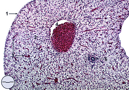
Figure 2. Cords of hepatocytes and central vein, transverse section
(Formalin, H&E, Bar = 84.8 µm). 1. liver
capsule; 2. cords of hepatocytes;
3. sinusoids containing red blood cells; 4. central vein; 5. bile
duct.
LIVER: Sinusoids and bile ducts
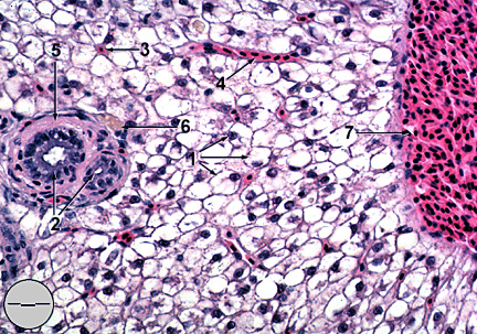
Figure 3. Sinusoids and bile ducts (Formalin, H&E, Bar
= 22.8 µm).
1. hepatocytes with glycogen vacuoles and eccentric nuclei; 2.
transverse
section of bile ducts; 3. transverse section of a sinusoid comprised
of six
hepatocytes surrounding a capillary; 4. sagittal section of a
sinusoid capillary;
5. connective tissue; 6. tissue macrophage; 7. central vein.
LIVER: Sinusoids and bile canuliculi
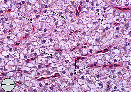
Figure 4. Sinusoids and bile canuliculi (Formalin,
H&E, Bar = 15.3 µm).
1. hepatocytes; 2. sagittal section through sinusoids; 3. bile
duct canuliculi.
PANCREAS: Exocrine and endocrine
tissue (a)
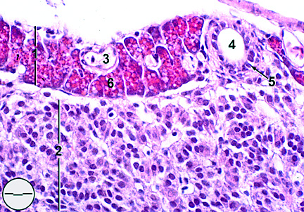
Figure 5. Exocrine and endocrine tissue (a) (Formalin, H&E,
Bar = 15.9 µm).
1. exocrine pancreatic tissue (acinar cells); 2. encapsulated
endocrine pancreatic
tissue (islets of Langerhans); 3. blood vessel; 4. pancreatic
duct; 5. cuboidal
epithelium; 6. zymogen granules.
PANCREAS: Exocrine and endocrine
tissue (b)
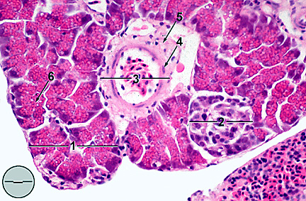
Figure 6. Pancreas, exocrine and endocrine tissue (b) (Formalin,
H&E,
Bar = 22.2 µm). 1. exocrine pancreatic tissue (acinar cells);
2. endocrine
pancreatic tissue (islets of Langerhans); 3. blood vessel; 4.
endothelium;
5. connective tissue; 6. zymogen granules.
[Back to Table of Contents]