RESPIRATORY SYSTEM Description
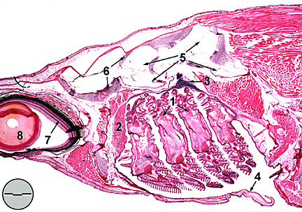
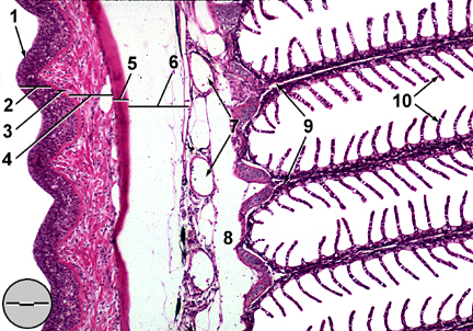
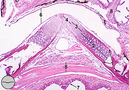
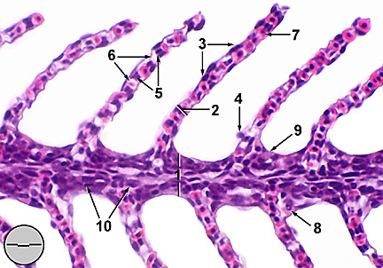
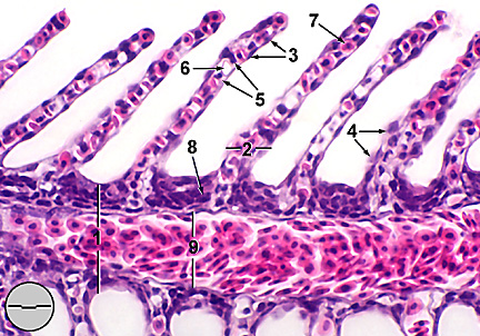
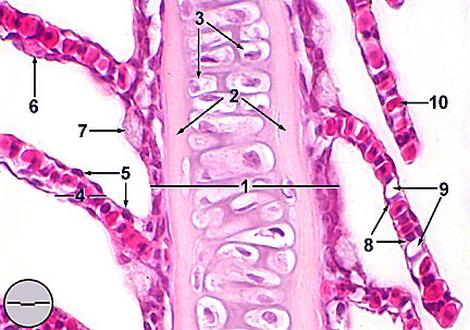
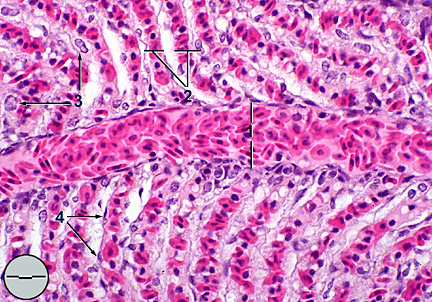
Gills Fathead minnow gills are comprised of four gill arches on either side and extending from floor to roof of the buccal cavity (Fig. 1). Two holobranchs (rows of filaments called primary lamellae) project from the posterior edge of each gill arch (Fig. 2). Anterior edges of the arches contain gill rakers which protect the fragile filaments and assist in food acquisition. Bony opercula protect the outside of the gills and regulate water movement across the filaments. Arches are supported by mixed bone (acellular and spongy) and cartilage with associated striated abductor and adductor muscles facilitating movement of gills to favorable respiratory positions (Fig. 3). Filaments have a central cartilaginous support (Fig. 6), afferent and efferent arterioles (Fig. 5), and a thin epithelial covering. This epithelium is contiguous with the covering of gill arches and the oral mucosa of the buccal cavity. Secondary lamellae originate on the superior and inferior surfaces of primary lamellae and are oriented perpendicular to the filaments. The thin epithelial covering of the secondary lamellae lies on a basement membrane supported by pillar cells. Spaces between pillar cells, called lacunae, connect afferent and efferent arterioles. The contractile pillar cells control the lacunar diameter thus regulating blood flow. Thin lamellar walls allow close proximity of blood within lacunae to the external environment. The direction of blood flow from afferent to efferent arterioles is opposite the direction of water flow over the lamellae. Short diffusion distances and counter-current blood flow promotes efficient exchange of oxygen and soluble metabolic wastes (e.g. carbon dioxide and ammonia). Other cell types found on primary and secondary lamellae include, melanocytes, lymphocytes, macrophages, endothelial cells, mucous cells, rodlet cells, and chloride cells. Vacuolated mucous cells, found primarily at the base of secondary lamellae, produce a thin mucus coating which protects against abrasion and bacterial infection and helps reduce drag. Due to their hyperosmotic state freshwater fish tend to passively lose ions and gain water through diffusion across the gills. Copious release of dilute urine, required to expel excess water, increases the loss of ions. Chloride cells, located between secondary lamellae on gill filaments (Fig. 4), provide the primary means for maintaining internal ionic homeostasis. Richly endowed with mitochondria and intricately laced with a tubular network of smooth endoplasmic reticulum, these cells actively transporting ions (e.g. Na+ and Cl-) against concentration gradients into the fish. Pseudobranch In teleosts pseudobranchiae occur bilaterally along the interior
of the opercula anterior to the first pair of gill arches (Fig. 1). While morphologically similar
to the gills, pseudobranchiae have a single row of filaments and
receive oxygen-rich blood from the first efferent arteries (and
are therefore unlikely to serve a respiratory function). In Cyprinidae,
including Pimephales promelas, the pseudobranch is completely
covered by the opercular epithelium. Lamellae, lacking any contact
with the external medium, are fused to each other forming a "glandular
pseudobranch" (Laurent and Dunel-Erb, 1984). The epithelium
is comprised predominantly of pseudobranchial cells which are
morphologically similar to chloride cells (but unique to the pseudobranch) (Fig. 7). Functions attributed
to the pseudobranch include; larval respiration before maturation
of gill arches, regulation of oxygen to the eyes, enzyme production
for use in the gas bladder, osmoregulation, and many others. In
a revue of pseudobranch morphology and function, Laurent and Dunel-Erb
(1984) found none of these explanations completely stisfactory,
concluding that a likely function "should be at least partly
sensory." These authors believe the pseudobranch, which is
richly innervated, functions primarily in maintaining blood pressure.
RESPIRATORY
SYSTEM: Buccal cavity, parasagittal section
RESPIRATORY
SYSTEM: Gill arch, sagittal section
RESPIRATORY
SYSTEM: Ceratobranchial bone of the gill arch,
RESPIRATORY
CHAPTER: Gill filament, sagittal section
RESPIRATORY
CHAPTER: Gill filament, sagittal section through
RESPIRATORY
CHAPTER: Gill filament, sagittal section through
RESPIRATORY
SYSTEM: Pseudobranch, sagittal section
|