RENAL SYSTEM Description
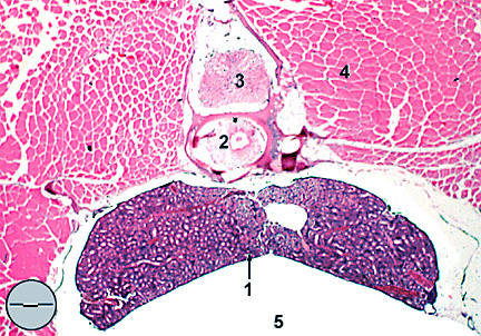
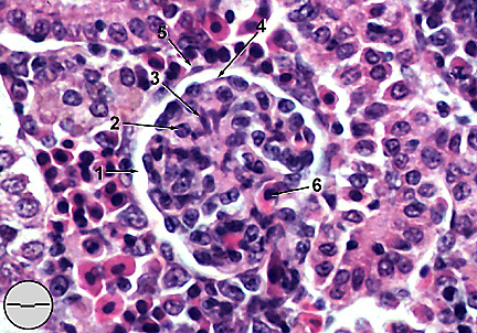
The fathead minnow kidney is located between the vertebral column and gas bladder extending longitudinally from cranium to anus (Fig. 1). The organ is divided into two portions, an anterior head kidney composed of hematopoeitic, lymphoid, and endocrine tissue, and a posterior trunk kidney composed of numerous nephrons surrounded by interstitial lymphoid tissue. Right and left sides of the trunk kidney are fused and form a deep saddle which occupies the space between the two chambers of the gas bladder. Posterior to this saddle the trunk kidney thins as it conforms to the curve of the gas bladder. The head kidney, separated into right and left sides, lies anterior to the saddle and penetrates into the cranium. Structure and function of the head kidney are discussed further in the sections on hematopoeitic and endocrine tissues. Body fluids of freshwater fish are higher in ionic concentration than the surrounding water, a conditions referred to as hyperosmotic. Maintaining such a concentration gradient requires removal and conservation of ions prior to excretion of "purified" water. This is accomplished in the kidney by filtration of water through glomerular nephrons each comprised of a renal corpuscle and renal tubule. The corpuscle consists of a vascular glomerulus enclosed within Bowman's capsule (Fig. 2). Outer parietal and inner visceral epithelia create "Bowman's space" which serves to isolate the glomerulus from the rest of the kidney. While fathead glomeruli are relatively small and avascular,
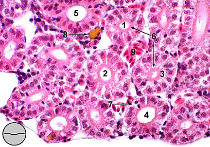
their renal tubules are structurally advanced possessing six cytologically
distinct regions (Fig. 3).
The neck region (1) is continuous with the parietal and visceral
epithelia of Bowman's capsule maintaining the isolation of the
glomerulus. In fatheads the neck region is long and thin with
a narrow lumen surrounded by ciliated cuboidal to low columnar
epithelial cells. Cytoplasm of these cells stains slightly basophilic.
The first proximal segment (2) is covered by tall columnar epithelial
cells with basal nuclei and slightly eosinophilic cytoplasm. An
apical "brush border" of microvilli protrudes deeply
into the lumen. The second proximal segment (3) has a still taller
columnar epithelium with more centrally located nuclei and a less
well developed brush border. Numerous large mitochondria cause
the cytoplasm to stain intensely eosinophylic. The intermediate
segment (4), long and well developed in the fathead minnow, has
a narrow lumen surrounded by cuboidal to short columnar epithelial
cells with an inconspicuous brush border. Staining is still strongly
eosinophilic. The distal segment (5) is lined with large, relatively
clear columnar epithelial cells. Nuclei are central and the brush
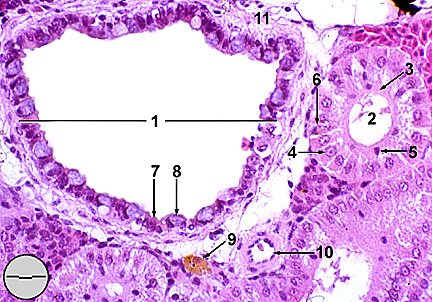
border is reduced or nonexistent. The initial collecting duct
(6) is larger in diameter than the preceding distal segment. Columnar
epithelium is lightly eosinophilic with basal nuclei and no brush
border. Subsequent collecting tubule segments increase in diameter
with their epithelium becoming pseudostratified and possessing
goblet cells. Larger collecting ducts incorporate layers of smooth
muscle and connective tissue (Fig.
4). Rodlet cells and intercalated cells (wandering leukocytes)
are common within the collecting duct epithelium.
RENAL
SYSTEM: Trunk kidney, transverse section
RENAL
SYSTEM: Glomerulus
RENAL
SYSTEM: Kidney tubules, transverse section (2)
RENAL
SYSTEM: Large collecting duct, transverse section
|