Necropsy: Completing the Necropsy


At the end of the necropsy, dispose of the carcass(es) in labeled, double plastic bags. Be sure to properly dispose of any "sharps," such as razor blades scalpels and needles. Be sure to have completely filled out all questions on your necropsy work sheet, including skin and gut scrape, results, gill biopsy results, hematocrit and serum protein levels, and all organ system observations. Make sure that all containers of fixative are securely closed and properly labeled.

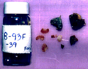
If there were several sample pieces of a particular organ which appeared normal, and one which had a remarkable observation, you might want to separate that piece of tissue by putting it into a labeled histology cassette inside the fixative container, or in a separate, labeled container. This way, you will be able to keep track of that tissue, through the histological processing, and on the glass microscope slide.

Roll your mouse cursor over the image.


After 24-48 hours of fixation, the tissues are ready to be "cut in." Sample small pieces of each preserve tissue specimen for dehydration and embedding in a block of paraffin wax. The next screen shows an example of the size of pieces to use for histological processing. Again, any specimen with a lesion should include both normal and abnormal areas.Depending on the size and curvature of the gill arches, they may or may not need to be cut. All cut specimen surfaces should be able to lay completely flat so that a good section may be processed from the paraffin block. Keep cut specimens small so as to reduce the number of slides needed. Note that the larger specimen in the upper right corner has a cut through an area containing both normal and potentially abnormal tissue. A single-edged razor is a good tool for cutting in specimens.

After the tissues are immersed in a series of xylene and alcohol for dehydration, they are embedded into paraffin blocks. These blocks are inserted into a holder on a microtome {definable button], and "thick" 6 micron sections of wax-containing tissue are sliced off. These slices are laid on glass microscope slides, paraffin melted off, and stained to help visualize the tissue structure. Hematoxylin (blue stain) and eosin (pink stain) is a common staining.After staining, a coverslip is placed over the slide with a mounting medium, and when dry, are ready for viewing under a microscope.

Back to Necropsy Menu