While fishes vary considerably in size, shape, and color, their external anatomy can be categorized using a single coherent system. The ability to describe very different fishes with this systematic vocabulary is useful in relating observations abut one species to another and to making comparison.
|
|
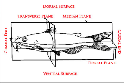
Before moving any further, you should review key terminology to orient yourself relative to the fish. Knowledge and use of "orientation" terminology will help you to describe your observations.
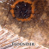
For example, you might make an observation something like the following: There are two 1-2 mm firm, white raised cysts, located one cm dorsal to the insertion of the pectoral fin.
Whenever possible, take a picture or make a sketch of the animal and draw the location of the lesion, along with any descriptive notes |
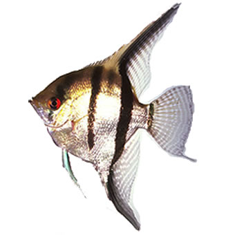
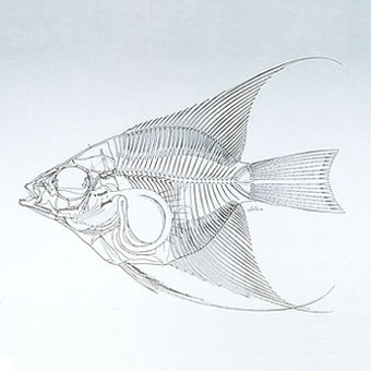
Angelfish
The images of the Anglefish below, show both external features and internal anatomical structures. These images show how closely the internal anatomy follows the external appearance. |
|
Angelfish |
Angelfish Skelston |
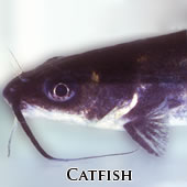
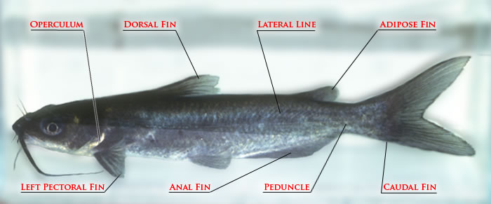
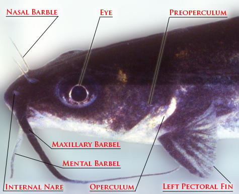
Catfish
The two photographs below show a freshwater Channel Catfish. The first is of the entire body with lines drawn to different external features. The second is of the head of the Catfish. Here you can see many of the smaller areas that are located between the pectoral fin and the internal nare. |
|
|
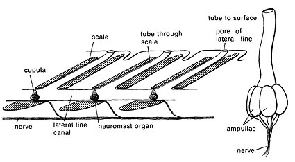
The image above points out an area on the fish known as the lateral line. To the left you can see a line drawing of this same area. Notice the nerves that run parallel to the lateral line canal. |
|
|
|
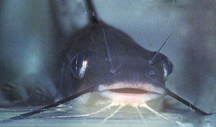
A frontal view of this channel catfish shows its pair of nasal barbels between the incurrent and excurrent openings of the nares. The long dark maxillary barbels and the medial and lateral mental (chin) barbels are also seen. |
|
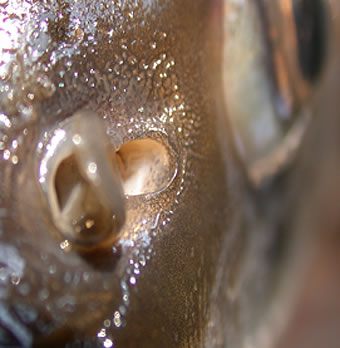
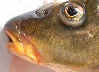
The Image to the left is of a fish nare located anterior to the mouth. The picture below is of a barble on the mouth of this Carp fish.
|
Skin
|
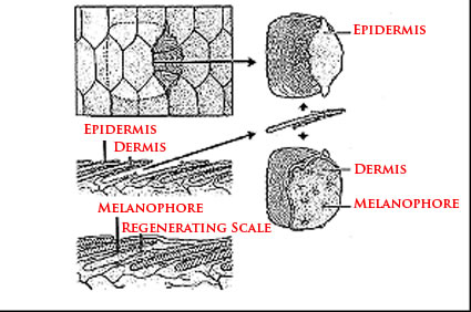
This section will take a closer look at the skin of fish. The diagram to he left and the key below will help you to correctly identify the different areas
|
|
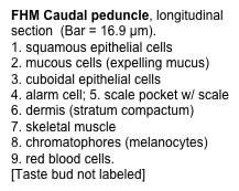
This line drawing of skin from a fish with scales (note that some fishes, like the channel catfish, lack scales) shows the overlapping nature of the squamation. It may also be evident that scales are in fact, of dermal origin, and have only a thin epidermal layer over them. Note the presence of melanophores within the dermis. |
|
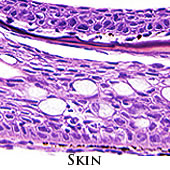
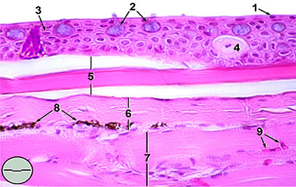
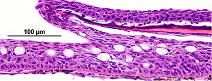
This is a histological cross-section of fish skin, stained with hematoxylin and eosin. Try and identify the overlying epidermis.
Melanophores (seen as small, black dots), and scales within scale pockets, are in the underlying dermis. |
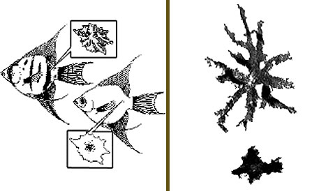
The above photo shows "light" and "dark-adapted" anglefish. The melanocyte visualized from this "dark-adapted" angelfish (upper-left) shows the melanin pigment distributed throughout the melanocyte, giving the cell an overall dark appearance. A melanocyte from the"light-adapted" angelfish (lower-right) shows the melanin pigment condenced in the central portion of the cell, giving the melanocyte an overall dark appearance.
|
The panel on the right shows two melanocytes taken from a skin scrape. The cell outlines are not seen, only the pigment granules are visible.. The melanocyte on the left has its pigment contracted, while the cell on the right has the pigment diffused within the cell, giving it an overall darker appearance. |
|
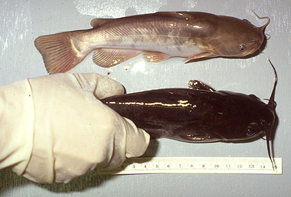
These two brown bullheads (Ictalurus nebulosis) were maintained in two different tanks: The upper fish was in a tank with a light background; the lower fish was kept in a tank with a dark background. Changes in coloration like this may take hours to days, depending on the intensity and quality of light, and the fish's state of health.
|
|
|

 External Anatomy .